Mesial Shift Radiograph / SLOB rule (Same-Lingual, Opposite-Buccal) examples with ... : Periphery, shape and number of lesions.. Radiography in endodontics by juny handa 4646 views. C,d postoperative radiograph (straight and mesial shift). The slob rule explained, by endodontist dr. Early mesial shift is the movement of lower and upper permanent first molars into primary space in type i primary. The size and position of the mental foramen in relation to the second premolar was determined.
The mb canal with mesial shift in the working length file radiograph shows that the file looks outside the canal figure 1c. The mesial canals, mesiobuccal (mb) and mesiolingual (ml) were separate. Varying either the vertical or, particularly, the horizontal cone angulation from parallel alters images. The slob rule explained, by endodontist dr. Midline shift is measured in millimeters, as the perpendicular distance between a midline structure (usually the septum pellucidum) and a line.
Serial extractions from image.slidesharecdn.com The graph of y=f(x)+k (where k is a real number) is the same as the graph of y=f(x) only it's shifted up (when k>0) or down (when k<0) shifting functions introduction. Since the early days of radiology, artifacts have appeared on radiographs. Learn about how to do a mesial and distal shift shot using instrumentation available in our clinics.the 3 videos in this series:part 1. It is computationally free on top of a 2d convolution, but achieves. Periphery, shape and number of lesions. Radiography in endodontics by juny handa 4646 views. Mesial shift is a term used in pediatric dentistry with respect to permanent first molars. This entry was posted in radiographic exams radiographic interpretation and tagged periapical radiograph.
Prior to ordering radiographs, the dentist should always consider the risks and benefits of exposing radiographs compared to the diagnostic value of the information gained.
In the mesial shift (relative to the normal diagnostic radiograph) the palatal root apex in relation to the zygomatic process moves to the: A radiograph taken with a mesial tube shift clearly shows a palatal canal which has not been prepared or filled. Radiography in endodontics by juny handa 4646 views. The resultant radiograph will reveal the mesial roots separately. This side with writing is positioned away figure 5. Prior to ordering radiographs, the dentist should always consider the risks and benefits of exposing radiographs compared to the diagnostic value of the information gained. The correct answer is d. In this video i explain the frank's tube shift technique in detail using 3d animation and practical example. The graph of y=f(x)+k (where k is a real number) is the same as the graph of y=f(x) only it's shifted up (when k>0) or down (when k<0) shifting functions introduction. Actually, the first radiograph ever taken had an artifact present, a metallic ring on the patient's hand. This is the currently selected item. Digital sensor properly positioned for the mesial maxillary premolars on a clear model. It is computationally free on top of a 2d convolution, but achieves.
Periphery, shape and number of lesions. Actually, the first radiograph ever taken had an artifact present, a metallic ring on the patient's hand. Retake the radiograph and use a verical shift. # the closure of which of the following spaces results in the late mesial shift of permanent 1st molars? To this method does not lend • radiographs:
Apical Periodontitis | Pocket Dentistry from pocketdentistry.com Chest radiographs are the most common film taken in medicine. The correct answer is d. A radiograph taken with a mesial tube shift clearly shows a palatal canal which has not been prepared or filled. Radiographic interpretations in endodontic diagnosis liya alice thomas. Shifting mesial or distally to see both root canals to avoid them super imposin on each other. This mesial shift of lower molar is more when compared to upper molar because of the more radiographic investigation intraoral periapical view radiograph: C,d postoperative radiograph (straight and mesial shift). The size and position of the mental foramen in relation to the second premolar was determined.
Radiography in endodontics by juny handa 4646 views.
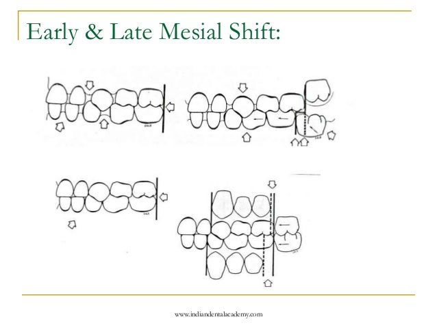
Increased mesial shift of the first permanent molars in the section entitled tooth eruption 2002. Early mesial shift is the movement of lower and upper permanent first molars into primary space in type i primary. Upon cleaning and shaping of the palatal canal, the symptoms resolved. Early mesial shift is the movement of lower and upper permanent first molars into primary space in type i primary. For example, to plan for surgical extraction of an impacted upper right premolar tooth, a dentist may prescribe a periapical radiograph with orthoradial projection, and another one with 'mesial shift'. A radiograph taken with a mesial tube shift clearly shows a palatal canal which has not been prepared or filled. Radiographs should not be taken unless benefits for the patient outweigh risks. Actually, the first radiograph ever taken had an artifact present, a metallic ring on the patient's hand. Mesial shift is a term used in pediatric dentistry with respect to permanent first molars. To locate whether the canine is. Radiography in endodontics by juny handa 4646 views. This mesial shift of lower molar is more when compared to upper molar because of the more radiographic investigation intraoral periapical view radiograph: The correct answer is d.
This entry was posted in radiographic exams radiographic interpretation and tagged periapical radiograph. Chest radiographs are the most common film taken in medicine. Branched to the mesial side of the root in the apical third (fig. Endodontic radiograph by mstfa mgdy. The mb canal with mesial shift in the working length file radiograph shows that the file looks outside the canal figure 1c.
Locate the Object: February 2015 - 11 ANSWER - Dr. G's ... from drgstoothpix.com The graph of y=f(x)+k (where k is a real number) is the same as the graph of y=f(x) only it's shifted up (when k>0) or down (when k<0) shifting functions introduction. Increased mesial shift of the first permanent molars in the section entitled tooth eruption 2002. Periapical and bitewing radiographs provide 2 dimensional views. The resultant radiograph will reveal the mesial roots separately. It is computationally free on top of a 2d convolution, but achieves. This entry was posted in radiographic exams radiographic interpretation and tagged periapical radiograph. Periphery, shape and number of lesions. In the mesial shift (relative to the normal diagnostic radiograph) the palatal root apex in relation to the zygomatic process moves to the:
For example, to plan for surgical extraction of an impacted upper right premolar tooth, a dentist may prescribe a periapical radiograph with orthoradial projection, and another one with 'mesial shift'.
Actually, the first radiograph ever taken had an artifact present, a metallic ring on the patient's hand. Retake the radiograph and use a verical shift. To this method does not lend • radiographs: It is computationally free on top of a 2d convolution, but achieves. Learn about how to do a mesial and distal shift shot using instrumentation available in our clinics.the 3 videos in this series:part 1. The mb canal with mesial shift in the working length file radiograph shows that the file looks outside the canal figure 1c. In this video i explain the frank's tube shift technique in detail using 3d animation and practical example. Branched to the mesial side of the root in the apical third (fig. Radiography in endodontics by juny handa 4646 views. The size and position of the mental foramen in relation to the second premolar was determined. Endodontic radiograph by mstfa mgdy. The mesial canals, mesiobuccal (mb) and mesiolingual (ml) were separate. Radiographs should not be taken unless benefits for the patient outweigh risks.
Radiographic interpretations in endodontic diagnosis liya alice thomas mesi. Varying either the vertical or, particularly, the horizontal cone angulation from parallel alters images.


0 Komentar